
The Plasma Membrane and the Fluid Mosaic Model

Let's take a look at a cell. One thing that you will find common among all cells is the cell membrane - whether it's an animal cell, a plant cell or a bacterial cell. While this layer may only be ten nanometers thick, it has an intricate molecular structure designed for efficient transport of material into and out of the cell. This property is known as selective permeability, the control of the passage of materials across the cell membrane.

The cell membrane is designed in a way that substances having certain properties are unable to enter or leave the cell (this movement across the cell boundary is known as transport). The fluid mosaic model describes how substances, mainly cholesterol, phospholipids and proteins, slide freely in the membrane.

Image result for fluid mosaic model

First, let's start with the phospholipid bilayer. A phospholipid a complex lipid with a "head" and a "tail". The head is made of one polar/hydrophilic phosphate group and a glycerol molecule. The tail is made two nonpolar/hydrophobic fatty acid chains. A fatty acid is a long chain of hydrocarbons (one carbon atom bonded to two hydrogen atoms) with a carboxyl group at one end. Glycerol is an alcohol with three carbon atoms.

Image result for phospholipid

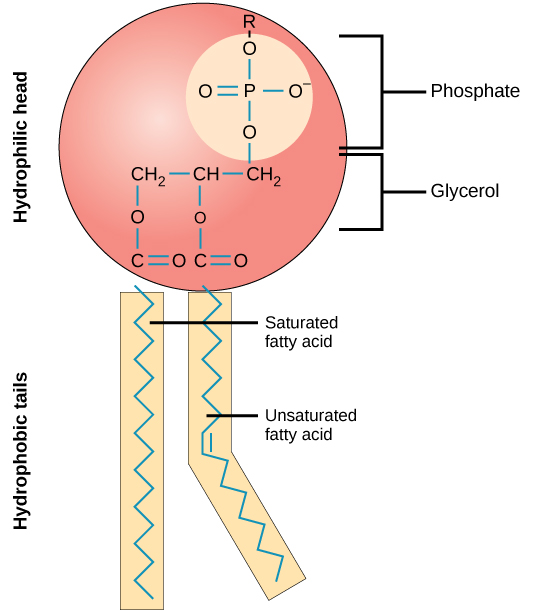
The phospholipids of the cell membrane are arranged in a bilayer (picture below). The hydrophilic (water-loving) head faces outward, where it has contact with the aqueous solution both inside and outside the cell. The hydrophobic (water-fearing) tail easily interacts with nonpolar molecules, but it interacts poorly with water. Because of this, the fatty acid tails are tucked away in the interior of the membrane to avoid any interactions with water. The chemical nature of the phospholipid bilayer only allows lipid-soluble molecules and some small molecules to freely pass across the membrane; ions and large polar molecules cannot.

Image result for phospholipid bilayer

                                                                                                                    ________
Image Credit:
(1) “Cell Membrane Detailed Diagram.” Wikimedia Commons, 31 Jan. 2007, commons.wikimedia.org/wiki/File:Cell_membrane_detailed_diagram_en.svg.

(2)  OpenStax, CNX. “Phospholipid.” Wikimedia Commons, 27 May 2016, commons.wikimedia.org/wiki/File:Figure_05_01_02.jpg.

(3) OpenStax. “Phospholipid Bilayer.” Wikimedia Commons, 18 May 2016, commons.wikimedia.org/wiki/File:0302_Phospholipid_Bilayer.jpg.

Comments

Popular Posts

Paul Broca and "Tan"

Paul Broca is one of the most influential neuroscientists best known for his patient, Tan. Well, his name wasn't really Tan. His name was Louis Victor Leborgne, and his incredibly unusual neurological disorder settled a debate about the location of language capabilities in the brain. In 1861, Leborgne approached Broca at the Bicetre Hospital to receive surgery for a leg infection. Leborgne had suffered from several medical conditions prior to his surgery; he had epilepsy at a young age and subsequently lost his ability to produce fluent speech. It was Leborgne's language disorder that really caught Broca's attention. Leborgne could think properly, but whenever he tried to communicate with Broca and verbalize his thoughts, all that came out of his mouth was the meaningless word "tan." For this reason, many scholars of neuroscience simply refer to Leborgne as "Tan". Broca realized that he could learn about our language capabilities by studying Leborg

Myelination

Neurons can transmit signals at astonishing speeds - some signals can travel as fast at 268 miles per hour! How are neurons capable of such speeds? Well, their axons, which transmit signals to other neurons, have a special covering known as the myelin sheath. Myelin, a lipid-rich substance, insulates the axon and increases the speed of signal transmission. As an action potential travels down the axon, some ions may cross the membrane and exit the cell. However, the presence of myelin prevents this escape. In the peripheral nervous system, myelin is found in the membranes of Schwann cells, a type of glial cell. Each Schwann cell forms one unit of myelin. In the central nervous system, oligodendrocytes, another type of glial cell, tightly wrap around the axon to form several layers of insulation. Each process of an oligodendrocyte can form one segment of myelin for several different cells. Myelin is not the only special feature of neurons that accelerates signal speeds. There are